Recently we have seen various cases of keratolytic disease that have showcased the various types of the disease there are. Here is a quick primer for horse owners on what it is and what you can and should do about it. Keratolytic disease or keratolysis of the zona alba has gone by several names over the years – hollow hoof, seedy toe, white line disease, while some of these names have remained popular they are not technically correct. Dr. Simon Curtis FWCF coined the term keratolysis of the zona alba which is the correct term from an anatomical and pathological standpoint.


Definition
Keratolytic disease is an infection of the hoof that is caused by a combination of an opportunistic bacteria and fungus that digest a portion of the hoof wall. As with many pathologies, this disease can be separated into multiple categories. In the thesis for her Fellowship, Sarah Logie FWCF described 2 types of keratolysis – Type 1 and Type 2. Type 1 – structural and Type 2 – Systemic. In structural type cases, as well as many systemic cases, the main area the pathogens affect is the Zona Alba, which is the bright white unpigmented portion of the hoof wall.
Anatomy
The structures we will talk about with keratolytic disease are the hoof wall, the white line, and the sole. We will start axially (towards the center of the foot) and work our way out. The sole is the horn that covers the majority of the bottom of the foot.
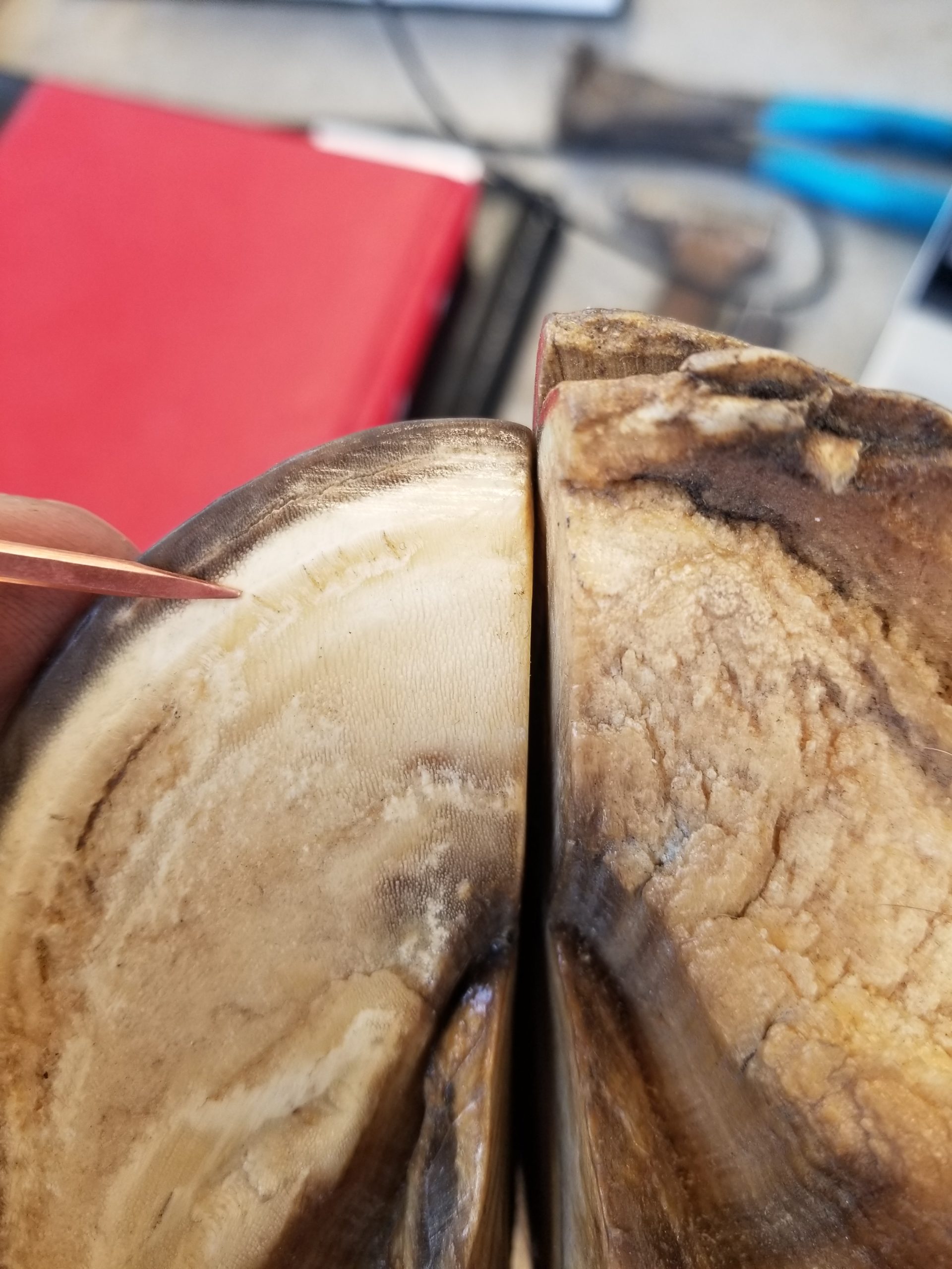
The white line is the structure that connects the sole to the hoof wall; it originates at the distal end (bottom) of the sensitive laminae. The hoof wall is made up of multiple layers. The majority of the mass of the hoof wall however is the stratum medium, which can be divided into two layers, the zona alba, which is the non pigmented inner layer, and the abaxial pigmented layer.
Etiology
The cause of keratolytic disease can be anything that allows microbes to invade the zona alba. In type 1 (structural) cases this will likely be a crack or separation due to excessive flare. Type 2 (systemic) is likely a genetic issue coupled with the environment.
Clinical signs
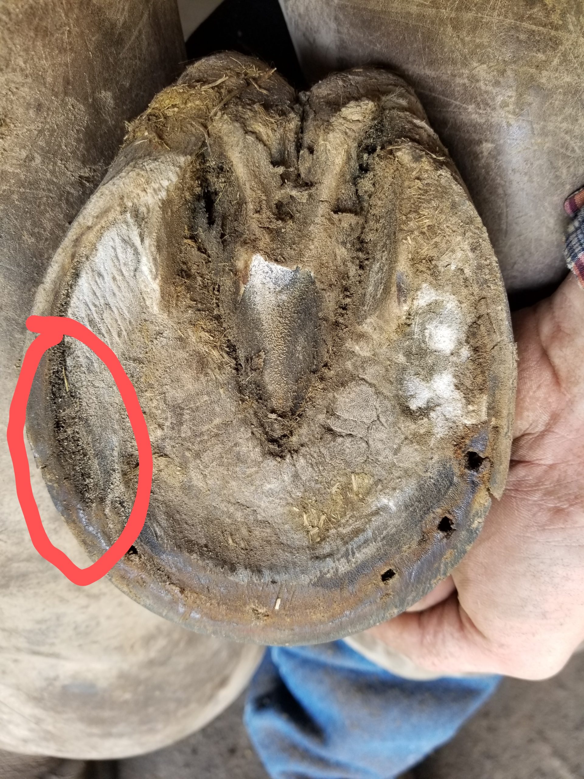
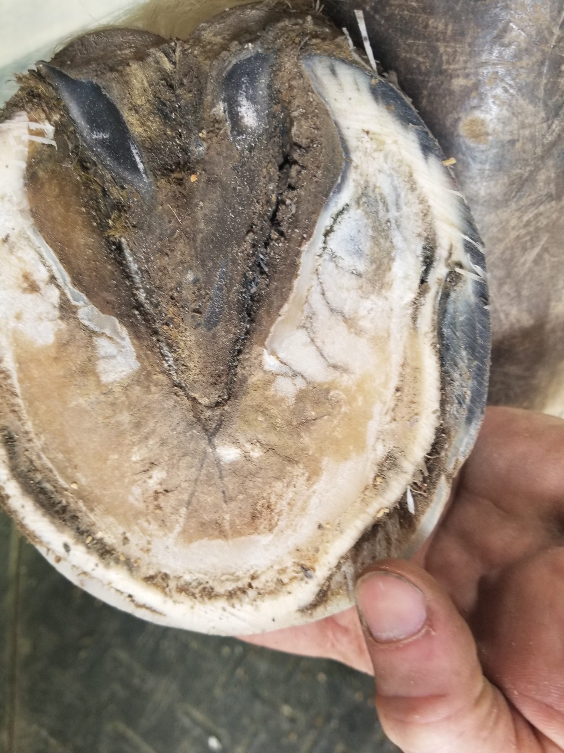
For type 1 and some type 2 cases the main clinical sign is a separation or cavity in the zona alba or non pigmented portion of the hoof wall. In some type 2 cases the separation may be present in the abaxial pigmented portion of the hoof wall; many horses with this superficial version of are overlooked with regard to microbial damage and are simply said to have poor or shelly feet.


Treatment
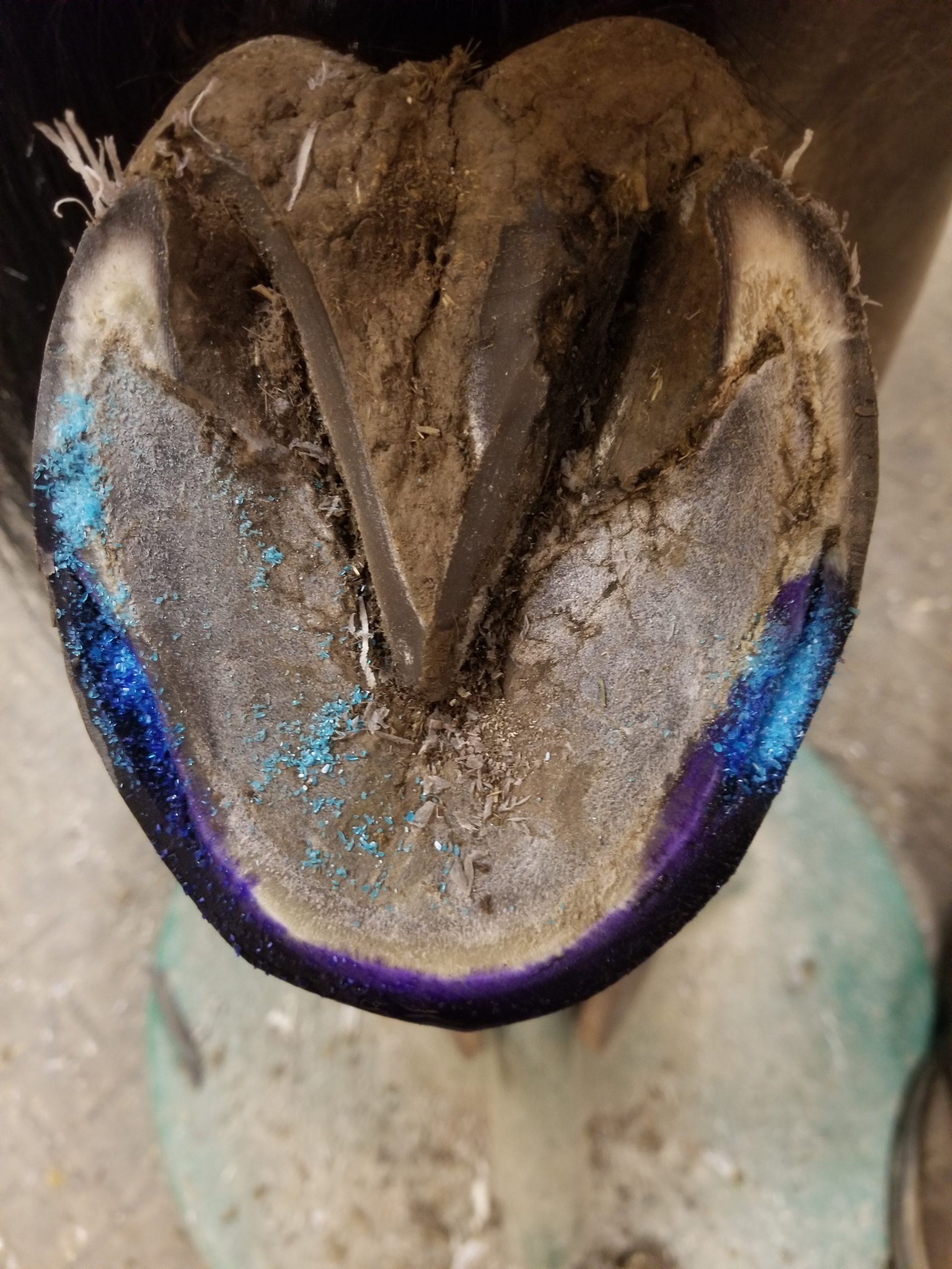
Horse owner treatment: Any separation or crack in the hoof wall should be treated with an antimicrobial in order to prevent the condition worsening. Treatment options include strong iodine, copper sulfate, or a thrush medication such as Thrush Buster.
A professional farrier and veterinarian should be consulted for more advanced cases as it may require a resection of the hoof wall.